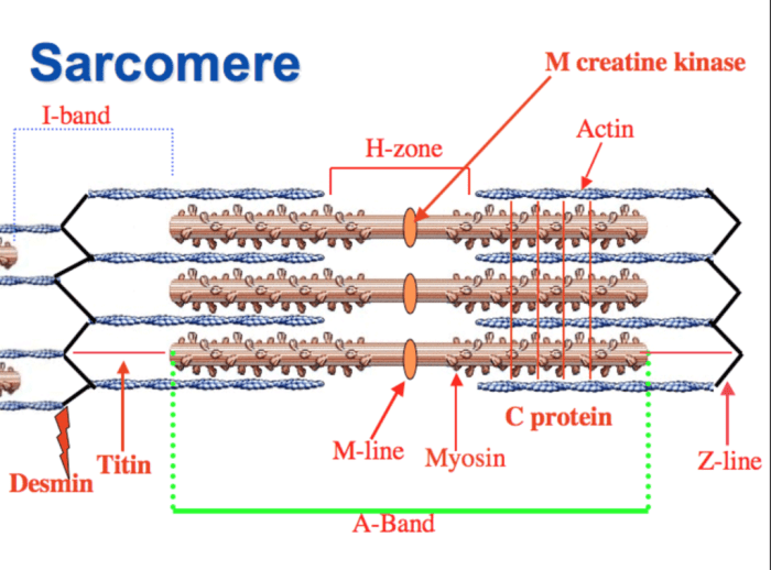
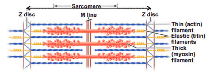
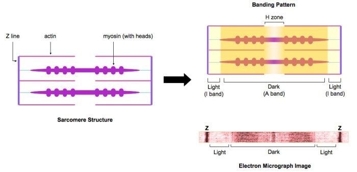
The drawing and photomicrograph below show a relaxed sarcomere – The drawing and photomicrograph below provide a captivating glimpse into the intricate structure of a relaxed sarcomere, the fundamental unit of muscle contraction. This detailed visual representation unveils the essential components and organization of myofilaments, setting the stage for an in-depth exploration of their role in muscle function.
Understanding the architecture of sarcomeres is paramount in comprehending muscle physiology and pathology. This comprehensive analysis delves into the distinct features of relaxed sarcomeres, comparing them to their contracted counterparts, and highlighting the critical role of calcium ions in mediating muscle contraction.
Understanding Sarcomere Structure in Skeletal Muscle

Sarcomeres are the fundamental contractile units of skeletal muscle, responsible for generating the force necessary for movement. Understanding their structure is crucial for comprehending muscle function and diagnosing muscle disorders.
Anatomy of a Relaxed Sarcomere
A relaxed sarcomere consists of the following components:
- A band:Contains thick myofilaments (myosin) and thin myofilaments (actin) overlapping in the central region.
- I band:Contains only thin myofilaments (actin), appearing as light bands on either side of the A band.
- Z line:A dense protein structure that anchors the thin myofilaments at the ends of the sarcomere.
- H zone:A narrow region within the A band where thick and thin myofilaments do not overlap.
Comparison of Relaxed and Contracted Sarcomeres
| Feature | Relaxed Sarcomere | Contracted Sarcomere |
|---|---|---|
| A band length | Unchanged | Shortens |
| I band length | Long | Shortens |
| Z line position | Fixed | Moves closer together |
| H zone width | Present | Disappears |
During contraction, calcium ions bind to troponin on the thin myofilaments, triggering a conformational change that allows the myosin heads to interact with actin, causing the sarcomere to shorten.
Role of the Drawing and Photomicrograph, The drawing and photomicrograph below show a relaxed sarcomere
The drawing provides a schematic representation of a relaxed sarcomere, highlighting the organization and relationship of its components. The photomicrograph, on the other hand, offers a real-world image of a sarcomere, showcasing its actual appearance under a microscope. Together, they complement each other by providing a comprehensive understanding of sarcomere structure.
Clinical Relevance
Understanding sarcomere structure is essential for diagnosing and treating muscle disorders. Mutations in genes encoding sarcomere proteins can lead to diseases such as hypertrophic cardiomyopathy (HCM) and dilated cardiomyopathy (DCM), which affect the heart muscle, and muscular dystrophy, which affects skeletal muscle.
FAQ Guide: The Drawing And Photomicrograph Below Show A Relaxed Sarcomere
What is the significance of understanding sarcomere structure?
Comprehending sarcomere structure is essential for deciphering muscle function and identifying the root causes of muscle disorders.
How do sarcomeres contribute to muscle contraction?
Sarcomeres are the fundamental units of muscle contraction, responsible for generating the force necessary for muscle movement.
What are the key differences between relaxed and contracted sarcomeres?
Relaxed sarcomeres exhibit distinct lengths and positions of the A band, I band, Z line, and H zone compared to contracted sarcomeres, reflecting changes in myofilament overlap.
What is the role of calcium ions in muscle contraction?
Calcium ions act as essential messengers, triggering the molecular events that lead to myofilament interaction and muscle contraction.

